
Open Journal of Clinical and
Medical Images
Research Article - Open Access, Volume 3
Reconstruction of posttraumatic facial defects
Cristina Stanescu1*; Camelia Tamas2; Georgiana-Daniela Stanescu3
1Faculty of Medicine and Farmacy ”Dunarea de jos” Galati, Romania.
2University of Medicine and Farmacy ”Gr. T. Popa” Iasi, Romania.
3Emergency County Hospital of Braila, Romania.
*Corresponding Author: Cristina Stanescu
Faculty of Medicine and Farmacy ”Dunarea de jos” Galati, Romania.
Email: dr.cristinastanescu@gmail.com
Received : Feb 09, 2023
Accepted : Mar 07, 2023
Published : Mar 14, 2023
Archived : www.jclinmedimages.org
Copyright : © Stanescu C (2023).
Abstract
The aim of surgical treatment is to restore both function and form and minimise the need for further interventions. Facial trauma is frequent associated with other injuries and a multidisciplinary approach must be required to establish the therapeutic plan (plastic surgeons, dermatologists, ophthalmologists and otorhinolaryngologists). Nasal trauma and fractures are some of the most frequent injuries in facial surgery practice. Fractures of the nose represent the third most common fracture of human body [4]. While nasal trauma and deformity are commonly recognised and treated, injuries to the adiacent structures (frontal process of the maxilla, the medial canthal tendon, orbit, nasal cartilages, nerves injuries) could be easily omitted with important consequences. The treatment of facial injuries is often delayed until more life-threatening problems have been solved, such as the establishment of an adequate permeable airway and hemodynamic stabilization. Isolated soft tissue wounds should be closed as soon as possible; early repairs have been associated with improving postoperative aesthetic results. Delays in surgical treatment increase the swelling of the soft tissue, obscuring landmarks and making primary closure more difficult with highly increased risk of infection. Successful reconstruction requires a thorough understanding of skin anatomy and physiology, a careful analysis of cutaneous defect and the consideration of multiple options for donor site. A local skin flap well planned for rapid reconstruction offers an adequate blood supply and a good color and texture of the skin. Each defect must be analyzed for depth, distortion of surrounding subunits and postoperative scarring.
Keywords: Facial trauma; Soft tisue injuries; Facial defects reconstruction; Local flaps.
Citation: Stanescu C, Tamas C, Stanescu GD. Reconstruction of posttraumatic facial defects. Open J Clin Med Images. 2023; 3(1): 1101.
Introduction
Facial trauma is frequent associated with other injuries. The presence of nasal trauma needs treatment strategies and algorithms to restore the nose to its original form and minimise the necesity for further surgery [4]. Injuries to the medial canthal region, orbit and naso-ethmoid region can lead to subsequent secondary deformities. The treatment of facial injuries is often delayed depending on associated lesions, such as the establishment of an adequate airway, hemodynamic stabilization. The severity of cranio-facial trauma is correlated with clinical expression [10]. Establishing airways permeability and hemodynamic stabilization are priorities. The incidence of complications including infection, asymmetry, hematoma and malocclusion were low in minor trauma [5]. The major cause of mortality is associated with systemic injuries and pulmonary infection [7]. Isolated soft tissue wounds should be closed as soon as possible; early repair of soft tissue and bone injuries have been associated with improved postoperative aesthetic results [8]. Delays in treatment can result in increased soft tissue swelling, obscuring landmarks and making primary closure more difficult with increasing risk of infection. Ideally, closure should occur within the first 8 hours after the injury when adequate haemostasis is required. Facial trauma often involves multiple aesthetic units and reconstruction is preferably planned for each unit such that incisions and local tissue used for advancement are within or along the border of the unit being reconstructed [6]. In the stabilized patient with complex facial injury immediate surgical treatment is indicated. Early reconstruction decreases the number of operations required without compromising the aesthetic or the functional outcomes [2].
Materials & Methods
A retrospective study includes 104 patients with craniofacial trauma treated at the Plastic Surgery Compartment at the Emergency County Hospital of Brăila between 2012 and 2019. Data regarding age, gender, etiology, anatomic site of facial fractures, nerve injuries associated, complications and therapeutic management were analyzed. The craniofacial injuries were identified after clinical examination and imaging investigations. Fractures of the facial skeleton were grouped as lower face (mandible), mid face (maxilla, nose, zygoma, and orbits) and upper face (frontal). Patients with sign and symptoms of systemic injuries to liver, kidney, bladder and bowels, hemothorax, pneumothorax, brain trauma and spinal cord injuries underwent suplimentary investigations (computed tomography, IRM).
Results and discussion
The immediate clinical examination is very important to determine the associate lesions. Five major mechanisms of injury were existed: traffic accidents (31%), sports (18%), violence (21%), work accidents (10%), activity of daily life (11%), animal bites (6%) and other causes (3%). Gender distribution showed an overall male-to-female ratio of 2 to 1, depending on the injury mechanism (a higher risk for males in facial bone fracture and for female in dental trauma during activities of daily life and violence). This high vulnerability of male gender for all types of trauma can be attributed to the facts that in our society male have more freedom to work outdoor, making them more vulnerable to accidents. Interpersonal violence is due to the increasing use of alcohol and drugs [1]. There was history of alcohol comsumption in 16% of patients. Loss of consciousness was the most common clinical symptom (54%) encountered in severe head trauma, followed by headache (41%) and require CT scan in all these cases. Other clinical features such as vomiting, nasal bleed and oral bleed had associated with cranio facial injuries. Isolated mandibular and nasal fractures are the most common facial bone fracture. All patients who sustain moderate or severe head ache have associated intracranial injuries reflecting the severity and complexity of craniofacial trauma [9]. The treatment of facial injuries is often delayed until more life-threatening problems have been solved, such as the establishment of an adequate permeable airway and hemodynamic stabilization [3].
Traumatic facial soft tissue injuries are commonly encountered in the emergency department by plastic surgeons and other providers. Although rarely life-threatening, the treatment of these injuries can be complex and may have significant impact on the patient’s facial function and aesthetics. This article provides a review of the relevant literature related to this topic and describes the authors’ approach to the evaluation and management of the patient with facial soft tissue injuries. Soft tissue injuries, whether isolated or in combination with other systemic injuries, are among the most common traumatic craniofacial injuries encountered by plastic surgery department. Smaller lacerations can be treated using local anesthesia, whereas larger injuries need general anesthesia. If significant contamination is present, wounds must be cleaned with antiseptic solution in the first 6 hours after injury. After cleansing, wound edges and devitalized tissues should be debrided. In cases, such as crush injuries, where the extent of injury is unclear, tissues can be loosely reapproximated, to obliterate dead spaces and to relieve tension on epidermis. The presence of contamination has not been associated with an increase in postoperative complications after early definitive repair of facial injuries with local flaps, therefore contamination should not be considered a contraindication to this treatment approach. Broad-spectrum antibiotic coverage is necessary in bite wounds and in patients with impaired wound healing due to smoking, alcoholism, diabetes, or other forms of immunodeficiencies.
Eyelid or periocular injuries could affected upper eyelid, lower eyelid, medial canthus, and lateral canthus. Simple eyelid lacerations should be closed in three layers: conjunctiva, tarsus and skin. For lower-lid lacerations, proper alignment minimizes the risk of ectropion. In upper-lid lacerations, the levator muscles should be preserved. Upper and lower eyelid full-thickness defects involving less than 25-30% of the eyelid length can be closed primarily. A lateral canthotomy and cantholysis can be used to relieve tension in larger defects. Defects involving up to 50% of the eyelid length can be closed using local advancement flaps (Figures 1,2).
Small scalp defects usually can be closed primarily. Larger defects require hair-bearing tissue for reconstruction with local rotation and advancement flaps. Skin grafting can be performed if the pericranium is intact [2]. Once the wound is closed, other options such as tissue expansion can be used to improve aesthetic outcome.
Most ear injuries can be trated under local anesthetic, with the exception of subtotal or complete avulsion, which requires immediate microsurgical treatment. Simple lacerations should be conservatively debrided to avoid unnecessary cartilage exposure. Small skin defects with an intact perichondrium can be repaired by skin grafting in most cases. If the perichondrium is not intact, the underlying cartilage can be resected, or a postauricular flap can be used to provide a vascularized bed for repair. Total ear avulsions should be immediately microsurgically replanted if donor and recipient vessels are available.
Nasal reconstruction have a great interest for plastic surgeons and a wide variety of reconstructive approaches have been developed to treat traumatic nasal injuries. Local and regional factors make reconstruction more complicated. The skin and soft tissues of the nasal tip and alar rim tend to be thick and relatively stiff, making primary closure of these regions more difficult. Partial-thickness defects over subunits with thinner skin can be closed using postauricular and supraclavicular full-thickness skin grafts. Local flaps such as dorsal nasal, cheek advancement, nasolabial and paramedian forehead flaps are used to repair soft tissue defects. Local flaps in facial reconstruction brings the best results (Figure 3,4,5). Tissue expanders can also be used to expand the available tissue without compromising the ability to close the donor site but require more aditional operations. Large defects require the use of distal free flaps and these patients have poor functional and aesthetic outcomes compared with those patients with other craniofacial injuries.
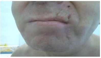
Many cheek wounds can be primary closure by direct suture due to the laxity of surrounding soft tissue. For large defects, local advancement, transposition, or regional flaps can be used and are generally preferred to skin grafting. In lip injury management primary closure in anatomical layers should be considered when less than 30% of the lip is involved (Figures 6,7). For defects of the central upper lip, primary closure may disrupt the normal anatomy of the columns of the philtrum. For wounds that cannot be primarily closed, the best method to achieve restoration is to use an Abbé flap to reconstruct the central vermillion without moving the commissure. Larger defects can be repaired using a variety of local advancement flaps. The reconstruction of soft tissue defects in facial injuries need appropriate surgical procedures for functional rehabilitation and aesthetic outcome.


Conclusions
In front of a patient with cranio-facial injuries presented at the Emergency Room, evaluation of vital parameters and immediate imaging investigations represent the first step in therapeutic management. In facial trauma, older persons are prone to bone fractures and soft tissue injuries while younger persons are more susceptible to dentoalveolar and facial bone lesions. Clinically, fronto-maxillary injuries may constitute a diagnostic problem, as their severity is not correlate with the patient's general condition. To establish a corect diagnosis, normal standard films and tomographs are required to identify fracture lines involving the base of the skull, nasal structure, periorbital region. A thorough, but focused, physical exam should be performed to assess soft tissue damage and determine the initial steps in therapeutic management. All wounds should be evaluated for size, depth, level of contamination, infection, integrity and viability of the wound edges. Evaluation and documentation of cranial nerve function, particularly the facial and trigeminal nerves, is critical if the patient is unconscious. Parotid (Stenson's) duct injuries should be suspected at any patient with facial trauma extending from the pretragal region to the middle half of the ipsilateral upper lip. In patients with a suspected parotid duct injury, the duct can be cannulated intraorally at the level of the second maxillary molar. The most serious complication associated with facial injuries is the occurrence of cerebrospinal rhinorrhoea. Other complications associated include intracranial haemorrhage, oculomotor dysfunction, obstruction of nasal airways, deficient lacrimal drainage, dental malocclusion, compression of optic nerve, facial nerve branches injuries etc. Factors such as time of presentation in relation to injury, degree of injury, and anatomical areas involved, play critical roles in determining the optimal management for surgical treatment. Scar revisions could be performed later after scar maturation.
Conflicts of interest statement: The authors declare no conflicts of interests.
References
- Le BT, Dierks EJ, Ueeck BA, Homer LD, Potter BF. Maxillofacial injuries associated with domestic violence. J Oral Maxillofac Surg. 2001; 59: 1277-1283.
- Seitz IA, Gottlieb LJ. Reconstruction of scalp and forehead defects. Clin Plast Surg. 2009; 36: 355-377.
- Sauaia A, Moore FA, Moore EE, Moser KS, Brennan R, et al. Epidemiology of trauma deaths: A reassessment. J Trauma. 1995; 38: 185-193.
- Parrett BM, Pribaz JJ. An algorithm for treatment of nasal defects. Clin Plast Surg. 2009; 36: 407-420.
- Kaur J, Bajwa SK, Kaur G, Singh A, Parmar SS, et al. Designing, managing and improving the operative and intensive care in polytrauma. J Emerg Trauma Shock. 2011; 4: 494-500urg. 2009; 36: 407-420.
- Stanley RB, Schwartz MS. Immediate reconstruction of contaminated central craniofacial injuries with free autogenous grafts. Laryngoscope. 1989; 99: 1011-1015.
- Gassner R, Tuli T, Hochl O, Rudisch A, Ulmer HJ. Cranio-maxillofacial trauma: A 10 year review of 9,543 cases with 21,067 injuries. J Craniomaxillofac Surg. 2003; 31: 51-61.
- Benzil Deborah L, Enrico R, Forcht DT, Patrick S, Jack BR, Neville KW. Early Single-Stage Repair of Complex Craniofacial Trauma. Neurosurgery. 1992; 30: 166-172.
- Cryer PC, Davidson L, Styles CP, Langley JD. Descriptive epidemiology of injury in the South East: Identifying priorities for action. Public Health. 1996; 110: 331-338.
- Derdyn C, Persing JA, Broaddus WC, Delashaw JB, Jane J, et al. Craniofacial trauma: An assessment of risk related to timing of surgery. Plast Reconstr Surg. 1990; 86: 238-245.